3 Distinctions and relationships of major groups of life
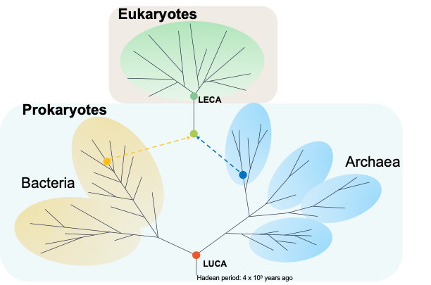
All cellular life forms are believed to have evolved from a common ancestor known as the Last Universal Common Ancestor (LUCA). The relationships among these life forms are illustrated by the “tree of life,” which has traditionally included three main domains: bacteria, archaea, and eukaryotes. Evolutionary biologists have been trying to determine LUCA’s position in this tree and to trace the origins of the Last Eukaryotic Common Ancestor (LECA). However, this task is complicated by the vast evolutionary timescales and the frequent exchange of genetic material between different microbial lineages. Recently, new data and techniques have significantly altered our understanding of the tree of life.
The basic blueprint of the first known living cells – LUCA
During the Hadean period, approximately 4.5 billion years ago, Earth was characterised by intense meteor bombardments, volcanic activity and collisions. Amidst these harsh conditions a small group of cells emerged, LUCA, sharing a common blueprint. LUCA possessed several key features: DNA in a double helix structure, RNA for transcription, proteins synthesised from a pool of 20 amino acids, ribosomes for protein assembly, a phospholipid membrane, ATP as an energy source, and Na/K ATPase to maintain a sodium/potassium gradient. LUCA contained 355 identifiable genes and thrived around deep ocean vents, which were both hot and acidic environments, making LUCA a thermophile and acidophile. One prevalent theory suggests that LUCA may have arrived on Earth via meteor bombardment, a common occurrence during the Hadean period.
The formation of LECA from LUCA
From the LUCA, two major groups (or domains) emerged, Bacteria and Archaea, collectively known as prokaryotes (Prokaryota). According to the endosymbiotic theory, an archaean ingested a bacterium, which then became an internally saprophytic organism. This event led to the development of a new type of living cell, forming a third domain called eukaryotes (Eukaryota). The first eukaryotic cells appeared around 1.2 billion years ago. This marks the emergence of LECA.
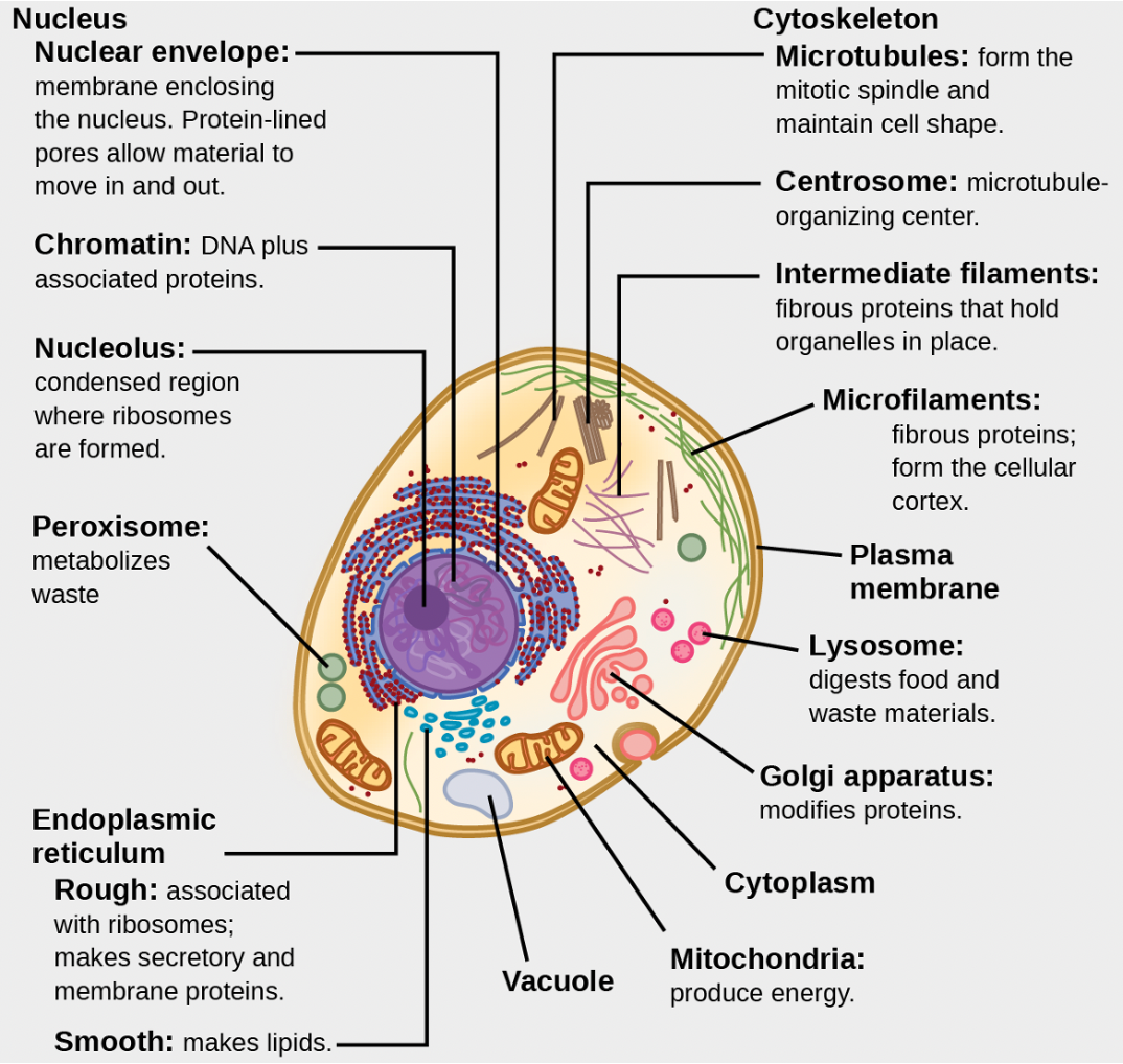
Eukaryotic cells, which are characteristic of all modern eukaryotes, share several fundamental features (Figure 1.13). These cells possess an endomembrane system, which includes structures such as the endoplasmic reticulum and the Golgi apparatus. They contain mitochondria or remnants of these organelles, essential for energy production. Eukaryotic cells also have a defined nucleus that contains their genetic material. Some eukaryotic cells have a primary cilium, a vestigial (redundant) form of flagellum. These components collectively define the complex architecture of eukaryotic cells.
Following an ice age and subsequent global melt, weathering and breakdown of rocks released essential minerals into the environment. During this period, oxygen concentrations were notably high, around 30%. Eukaryotes began forming colonial organisms, which are collectives of similar cells without specialised functions. Approximately 650 million years ago, multicellular organisms appeared. From these early multicellular eukaryotes, four major groups emerged: metazoans (animals), fungi, protists, and plants.
Animal cells differ significantly from single-cell eukaryotes in several ways. They develop complex intercellular junctions, allowing them to adhere to one another and facilitate intercellular communication. Animal cells exhibit massive gene duplication and possess an expanded genetic code, with over 50,000 genes. This genetic complexity enables specialisation of cells, which requires the ability to selectively silence genes, a process known as function inhibition. Notably, cancer represents a loss of this gene silencing ability. The earliest animals, such as molluscs and arthropods, exemplify these advanced cellular characteristics.
Where are LUCA and LECA positioned on the evolutionary tree of life?
The tree of life contains three major branches—bacteria, archaea, and eukaryotes (Figure 1.14). It is believed that eukaryotes originated when an archaeal cell absorbed a bacterial endosymbiont (endosymbiotic formation), forming a proto-eukaryote with a proto-mitochondrion. Gene duplications in LECA support this early mitochondrial origin, showing bacterial genes were copied into the archaeal host genome. Eukaryotic genomes have more bacterial genes than archaeal genes, with bacteria contributing 53% of genes in non-photosynthetic eukaryotes and 61% in photosynthetic ones.
‘Ontogeny Recapitulates Phylogeny’ suggests that the development of an organism’s organs (ontogeny) mirrors the evolutionary history of its species (phylogeny). Phylogeny refers to the relatedness of animals throughout Earth’s fossil record, organised from the simplest to the most complex forms. Simpler forms tend to predate more complex ones. By charting the progression from simple cells like archaea and bacteria to increasingly complex multicellular organisms, we can define a “tree” of relationships. This increasing complexity is reflected in the ontogeny of animals, which develop in utero from a single egg cell into complex organisms.
