11.4 The Stomach
Although a minimal amount of carbohydrate digestion occurs in the mouth, chemical digestion really gets underway in the stomach. An expansion of the gastrointestinal tract that lies immediately inferior to the oesophagus, the stomach links the oesophagus to the first part of the small intestine (the duodenum) and is fixed in place at its oesophageal and duodenal ends. In between, however, it can be a highly active structure, contracting and continually changing position and size. These contractions provide mechanical assistance to digestion.
An important function of the stomach is to serve as a temporary holding chamber. The stomach plays several important roles in chemical digestion, including the continued digestion of carbohydrates and the initial digestion of proteins and triglycerides. Little if any nutrient absorption occurs in the stomach.
Structure
There are four main regions in the stomach: the cardia, fundus, body, and pylorus (Figure 11.12). The cardia (or cardiac region) is the point where the oesophagus connects to the stomach and through which food passes into the stomach. Located inferior to the diaphragm, above and to the left of the cardia, is the dome-shaped fundus. Below the fundus is the body, the main part of the stomach. The funnel-shaped pylorus connects the stomach to the duodenum. The wider end of the funnel, the pyloric antrum, connects to the body of the stomach. The narrower end is called the pyloric canal, which connects to the duodenum. The smooth muscle pyloric sphincter is located at this latter point of connection and controls stomach emptying. In the absence of food, the stomach deflates inward, and its mucosa and submucosa fall into a large fold called a ruga (rugae – plural)

The convex lateral surface of the stomach is called the greater curvature; the concave medial border is the lesser curvature. The stomach is held in place by the lesser omentum, which extends from the liver to the lesser curvature, and the greater omentum, which runs from the greater curvature to the posterior abdominal wall.
 Case study
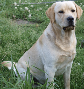
A 2-year-old Labrador Retriever, Max, presented with persistent vomiting and lethargy. Diagnostic imaging revealed a gastric foreign body causing mechanical obstruction. Labradors are known for indiscriminate eating, and in this case, a sock was identified as the obstructing object! A laparotomy was performed under anaesthesia to remove the sock! and Max recovered well with supportive care.
Histology
The wall of the stomach is made of the same four layers as most of the rest of the gastrointestinal tract, but with adaptations to the mucosa and muscularis for the unique functions of this organ. In addition to the typical circular and longitudinal smooth muscle layers, the muscularis has an inner oblique smooth muscle layer (Figure 11.13). As a result, in addition to moving food through the tract, the stomach can vigorously churn food, mechanically breaking it down into smaller particles.

The stomach mucosa’s epithelial lining consists only of surface mucus cells, which secrete a protective coat of alkaline mucus. A vast number of gastric pits dot the surface of the epithelium, giving it the appearance of a well-used pincushion, and mark the entry to each gastric gland, which secretes a complex digestive fluid referred to as gastric juice.
Although the walls of the gastric pits are made up primarily of mucus cells, the gastric glands are made up of diverse types of cells. The glands of the cardia and pylorus are composed primarily of mucus-secreting cells. Cells that make up the pyloric antrum secrete mucus and several hormones, including the majority of the stimulatory hormone, gastrin. The much larger glands of the fundus and body of the stomach, the site of most chemical digestion, produce most of the gastric secretions. These glands are made up of a variety of secretory cells. These include parietal cells, chief cells, mucous neck cells, and enteroendocrine cells.
Parietal cells—Located primarily in the middle region of the gastric glands are parietal cells, which are among the most highly differentiated of the body’s epithelial cells. These large cells produce both hydrochloric acid (HCl) and intrinsic factor. HCl is responsible for the high acidity (pH 1.5 to 3.5) of the stomach contents and is needed to activate the protein-digesting enzyme, pepsin. The acidity also kills much of the bacteria you ingest with food and helps to denature proteins, making them more available for enzymatic digestion. Intrinsic factor is a glycoprotein necessary for the absorption of vitamin B12 in the small intestine.
Chief cells—Located primarily in the basal regions of gastric glands are chief cells, which secrete pepsinogen, the inactive proenzyme form of pepsin. HCl is necessary for the conversion of pepsinogen to pepsin.
Mucous neck cells—Gastric glands in the upper part of the stomach contain mucous neck cells that secrete thin, acidic mucus that is much different from the mucus secreted by the goblet cells of the surface epithelium. The role of this mucus is not currently known.
Enteroendocrine cells—Finally, enteroendocrine cells found in the gastric glands secrete various hormones into the interstitial fluid of the lamina propria. These include gastrin, which is released mainly by enteroendocrine G cells. Table 11.6 describes the digestive functions of important hormones secreted by the stomach.
Table 11.6 Hormones secreted by stomach
| Hormone | Production site | Production stimulus | Target organ | Action |
|---|---|---|---|---|
| Gastrin | Stomach mucosa, mainly G cells of the pyloric antrum | Presence of peptides and amino acids in stomach | Stomach | Increases secretion by gastric glands; promotes gastric emptying |
| Stomach mucosa, mainly G cells of the pyloric antrum | Presence of peptides and amino acids in stomach | Ileocaecal valve | Relaxes valve | |
| Stomach mucosa, mainly G cells of the pyloric antrum | Presence of peptides and amino acids in stomach | Large intestine | Triggers mass movements | |
| Ghrelin | Stomach mucosa, mainly fundus | Fasting state (levels increase just prior to meals) | Hypothalamus | Regulates food intake, primarily by stimulating hunger and satiety |
| Histamine | Stomach mucosa | Presence of food in stomach | Stomach | Stimulates parietal cells to release |
| Somatostatin | Mucosa of stomach, especially pyloric antrum; also duodenum | Presence of food in the stomach; sympathetic axon stimulation | Stomach | Restricts all gastric secretions, gastric motility, and emptying |
| Mucosa of stomach, especially pyloric antrum; also duodenum | Presence of food in stomach; sympathetic axon stimulation | Pancreas | Restricts pancreatic secretions | |
| Mucosa of stomach, especially pyloric antrum; also duodenum | Presence of food in the stomach; sympathetic axon stimulation | Small intestine | Reduces intestinal absorption by reducing blood flow |
Gastric Secretion
The secretion of gastric juice is controlled by both nerves and hormones. Stimuli in the brain, stomach, and small intestine activate or inhibit gastric juice production. This is why the three phases of gastric secretion are called the cephalic, gastric, and intestinal phases (Figure 11.14). However, once gastric secretion begins, all three phases can occur simultaneously.

The cephalic phase (reflex phase) of gastric secretion, which is brief, takes place before food enters the stomach. The smell, taste, sight, or thought of food triggers this phase. Depression and loss of appetite can suppress the cephalic reflex.
The gastric phase of secretion lasts 3 to 4 hours and is set in motion by local neural and hormonal mechanisms triggered by the entry of food into the stomach. When food reaches the stomach, it creates distention that activates the stretch receptors. This stimulates parasympathetic neurons to release acetylcholine, which then provokes increased secretion of gastric secretion (‘juice’). Partially digested proteins, caffeine, and rising pH stimulate the release of gastrin from enteroendocrine G cells, which in turn induces parietal cells to increase their production of HCl, which is needed to create an acidic environment for the conversion of pepsinogen to pepsin, and protein digestion. Additionally, the release of gastrin activates vigorous smooth muscle contractions. However, it should be noted that the stomach does have a natural means of avoiding excessive acid secretion and potential heartburn. Whenever pH levels drop too low, cells in the stomach react by suspending HCl secretion and increasing mucous secretions.
The intestinal phase of gastric secretion has both excitatory and inhibitory elements. The duodenum has a key role in regulating the stomach and its emptying. When partially digested food fills the duodenum, intestinal mucosal cells release a hormone called intestinal (enteric) gastrin, which further excites gastric secretions. This stimulatory activity is brief, however, because when the intestine distends with chyme, the enterogastric reflex inhibits secretion. One of the effects of this reflex is to close the pyloric sphincter, which blocks additional acidic chyme from entering the duodenum.
The Mucosal Barrier
The mucosa of the stomach is exposed to the highly corrosive acidity of gastric juice. Gastric enzymes that can digest protein can also digest the stomach itself. The stomach is protected from self-digestion by the mucosal barrier. This barrier has several components. First, the stomach wall is covered by a thick coating of bicarbonate-rich mucus. This mucus forms a physical barrier, and its bicarbonate ions neutralise acid. Second, the epithelial cells of the stomach’s mucosa meet at tight junctions, which block gastric juice from penetrating the underlying tissue layers. Finally, stem cells located where gastric glands join the gastric pits quickly replace damaged epithelial mucosal cells, when the epithelial cells are shed. In fact, the surface epithelium of the stomach is completely replaced every 3 to 6 days.
Case study
A 4-year-old Schnauzer presented with chronic vomiting, intermittent anorexia, and signs of abdominal discomfort. Physical examination revealed mild dehydration and weight loss. Bloodwork showed elevated gastric enzymes, and gastroscopy revealed mucosal inflammation in the stomach lining. Biopsy samples confirmed the presence of Helicobacter pylori, a spiral-shaped bacterium known to colonise the gastric mucosa and cause gastritis and peptic ulcers. The dog was treated with a combination of antibiotics (amoxicillin and metronidazole) and a proton pump inhibitor to reduce gastric acidity. Clinical signs resolved within two weeks.
Miniature Schnauzer by Canarian via Wikimedia Commons, CC BY SA 3.0
Digestive Functions of the Stomach
The stomach participates in all the digestive activities except for ingestion and defaecation. Although almost all absorption takes place in the small intestine, the stomach does absorb some non-polar substances, such as alcohol and aspirin.
Mechanical Digestion
Within a few moments after food after enters the stomach, mixing waves begin to occur at intervals of approximately 20 seconds. A mixing wave is a unique type of peristalsis that mixes and softens the food with gastric secretions to create chyme. The initial mixing waves are gentle, but these are followed by more intense waves, starting at the body of the stomach and increasing in force as they reach the pylorus.
The pylorus, which holds around 30 mL of chyme, acts as a filter, permitting only liquids and small food particles to pass through the mostly, but not fully, closed pyloric sphincter. In a process called gastric emptying, rhythmic mixing waves force about 3 mL of chyme at a time through the pyloric sphincter and into the duodenum. Release of a greater amount of chyme at one time would overwhelm the capacity of the small intestine to handle it. The rest of the chyme is pushed back into the body of the stomach, where it continues mixing. This process is repeated when the next mixing waves force more chyme into the duodenum.
Gastric emptying is regulated by both the stomach and the duodenum. The presence of chyme in the duodenum activates receptors that inhibit gastric secretion. This prevents additional chyme from being released by the stomach before the duodenum is ready to process it.
Chemical Digestion
The fundus plays a key role, because it stores both undigested food and gases that are released during the process of chemical digestion. Food may sit in the fundus of the stomach for a while before being mixed with the chyme. While the food is in the fundus, the digestive activities of salivary amylase continue until the food begins mixing with the acidic chyme. Ultimately, mixing waves incorporate this food with the chyme, the acidity of which inactivates salivary amylase and activates lingual lipase. Lingual lipase then begins breaking down triglycerides into free fatty acids, and mono- and diglycerides.
The breakdown of protein begins in the stomach through the actions of HCl and the enzyme pepsin. During infancy, gastric glands also produce rennin, an enzyme that helps digest milk protein.
Its numerous digestive functions notwithstanding, there is only one stomach function necessary to life: the production of intrinsic factor. The intestinal absorption of vitamin B12, which is necessary for both the production of mature red blood cells and normal neurological functioning, cannot occur without intrinsic factor.
The Rumen
In ruminant herbivores like cows, sheep and antelopes the stomach is highly modified to act as a “fermentation vat”. It is divided into four parts. The largest part is called the rumen. In the cow it occupies the entire left half of the abdominal cavity and can hold up to 270 litres. The reticulum is much smaller and has a honeycomb of raised folds on its inner surface. In the camel the reticulum is further modified to store water. The next part is called the omasum with a folded inner surface. Camels have no omasum. The final compartment is called the abomasum. This is the ‘true’ stomach where muscular walls churn the food and gastric juice is secreted (Figure 11.15).

Figure 11.15 The Rumen.
Case study
Ruminants swallow the grass they graze almost without chewing and it passes down the oesophagus to the rumen and reticulum. Here liquid is added and the muscular walls churn the food. These chambers provide the main fermentation vat of the ruminant stomach. Here bacteria and single-celled animals start to act on the cellulose plant cell walls. These organisms break down the cellulose to smaller molecules that are absorbed to provide the cow or sheep with energy. In the process, the gases methane and carbon dioxide are produced. These cause the “burps” you may hear cows and sheep making.
Not only do the micro-organisms break down the cellulose but they also produce the vitamins E, B and K for use by the animal. Their digested bodies provide the ruminant with the majority of its protein requirements.
In the wild grazing is a dangerous activity as it exposes the herbivore to predators. They crop the grass as quickly as possible and then when the animal is in a safer place the food in the rumen can be regurgitated to be chewed at the animal’s leisure. This is ‘chewing the cud’ or rumination. The finely ground food may be returned to the rumen for further work by the microorganisms or, if the particles are small enough, it will pass down a special groove in the wall of the oesophagus straight into the omasum. Here the contents are kneaded and water is absorbed before they pass to the abomasum. The abomasum acts as a “proper” stomach and gastric juice is secreted to digest the protein.
Case study
A 2-week-old Fresian calf presented with bloating and mild diarrhoea. On examination, the calf was alert but had a distended abdomen. The farmer reported feeding milk via a bucket rather than a nipple. Normally, milk bypasses the rumen via the oesophageal groove and enters the abomasum for digestion. However, in this case, improper feeding technique failed to stimulate groove closure, allowing milk to enter the rumen. Fermentation of milk in the rumen led to gas production and digestive upset. Treatment included withholding milk temporarily, administering anti-bloat medication, and switching to bottle feeding to encourage proper oesophageal groove function. This case highlights the importance of feeding method in neonatal ruminants and the anatomical adaptations that support early digestion before full rumen development.

Holstein Friesian Calf by Ranjithkumar Murugesan via Wikimedia Commons, Public Domain
Section Review
The stomach participates in all digestive activities except ingestion and defaecation. It vigorously churns food. It secretes gastric secretions that break down food and absorbs certain drugs, including aspirin and some alcohol. The stomach begins the digestion of protein and continues the digestion of carbohydrates and fats. It stores food as an acidic liquid called chyme and releases it gradually into the small intestine through the pyloric sphincter.
Review Questions
Critical Thinking Questions
Click the drop down below to review the terms learned from this chapter.
